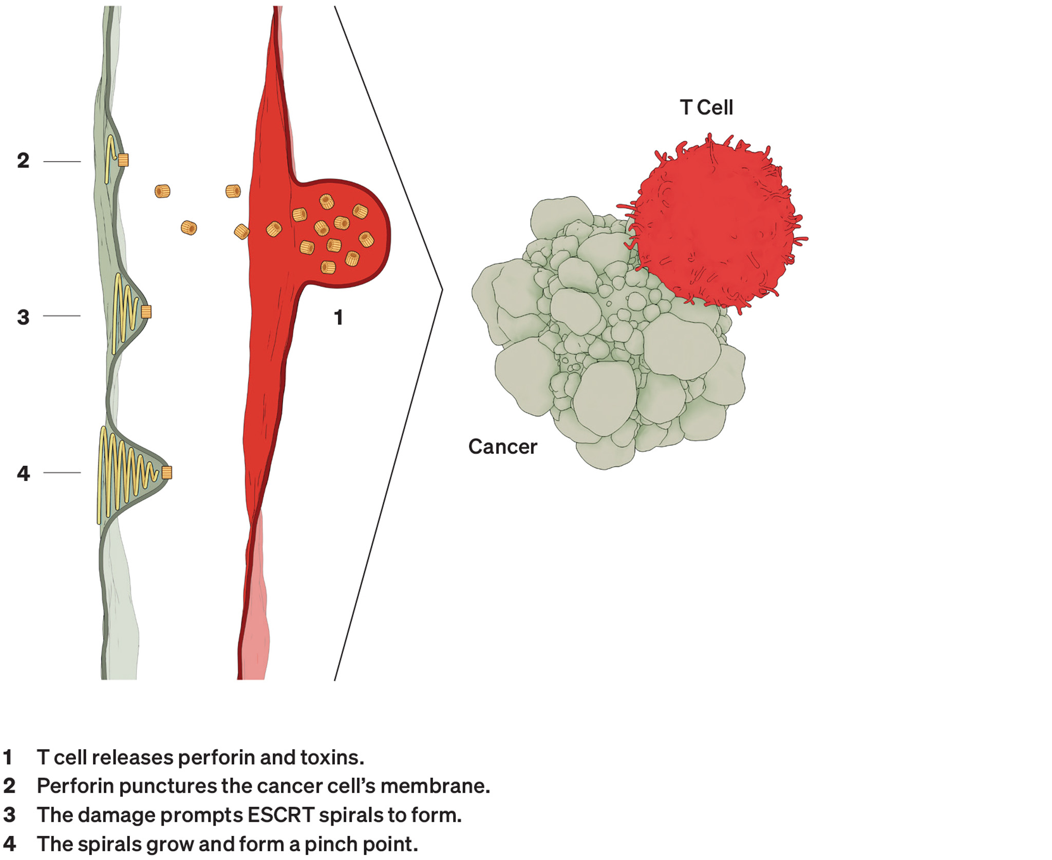
Microscopic image of a yellow arc-shaped membrane.
Illustrations: Anatomy Blue

Caught on Camera

Features

By tracing the contours of the proteins embedded in the membrane of cells, recordings made in Dr. Simon Scheuring’s lab reveal how these elusive molecules get their jobs done — for good and ill.

By Wynne Parry

Pushing Boundaries

Like a person or a country, a cell has boundaries. Yet a cell’s thin, skin-like border doesn’t just define a living space. It also hosts an abundance of life-sustaining activity. Certain molecules embedded within the double-layered membrane control access to the cell’s interior, while others sense stimuli such as light, hormones or mechanical force. Still others package cargo, interact with its skeleton and much more.

To understand how membrane proteins carry out these tasks, one lab at Weill Cornell Medicine captures them in action on video.

Using an advanced imaging technique known as high-speed atomic force microscopy (HS-AFM), Dr. Simon Scheuring, a professor of physiology and biophysics, and his colleagues make the molecular equivalent of documentaries, capturing the behavior of these elusive proteins as it plays out across minuscule fractions of a second within their natural habitat, the membrane.

“When you look at single molecules with HS-AFM, it’s like you’re making movies of them going about their business, just like people walking in the street,” Dr. Scheuring says.

The insight his group gleans into these routines casts new light on fundamental processes crucial to health and implicated in disease.

Real World, Real Time

When seeking to see the form and function of proteins, scientists have a handful of options. With techniques known as X-ray crystallography and cryo-electron microscopy, they can create highly detailed snapshots of proteins’ architecture. But to make these images, they must remove the proteins from the membrane and turn them into crystals for crystallography, or flash freeze them for cryo-electron microscopy. To see how a protein moves, they can track fluorescent tags they have applied to the protein, but not the movement of the protein itself.

By comparison, HS-AFM allows researchers to see the proteins themselves at work within the membrane. This approach is uniquely suited to catching the fleeting, and sometimes unexpected, transitions these molecules can undergo.

Although HS-AFM produces imagery, it functions more like the sense of touch than sight. A finely tipped probe, measuring only a few atoms thick at its point, moves over a sample. The tip traces the contours of the surface below it, recording changes in the molecules’ topography as they move.

Events unfold quickly at the molecular scale, so HS-AFM must work rapidly to catch them. Dr. Scheuring and his colleagues are continuing to push the limits on its speed. At present, the fastest version can capture a channel protein opening or closing over approximately 50 millionths of a second.


High-speed Atomic Force MicroscopyIllustration of imaging technique in which a laser diode bounces off of a molecule sample.

This imaging technique uses a fine tip to trace the surface of molecules. A laser reports the position of the tip.

Dr. Scheuring’s lab has used HS-AFM to study many membrane structures, including two on opposing sides in cancer: the immune protein perforin, which punctures cells that pose a threat, and ESCRT, a set of proteins that forms spirals and heals wounds, among other functions.

Perforin

Microscopic image of a yellow arc shaped membrane.

HS-AFM videos have shown that a break in a perforin ring triggers its segments to rotate and unfurl downward, attacking the membrane below. Typically, this transition produces an arc, not a full ring, above the hole it has punctured.

ESCRT

Microscopic image of yellow proteins in a spiral

Dr. Scheuring’s lab has explored how this set of proteins forms rings, then spirals, which accumulate mechanical force like a spring and change the shape of the membrane, a necessary step for many processes within a cell.

Co-opted by Cancer

Dr. Scheuring’s lab has applied HS-AFM to numerous membrane protein structures, including two that other research suggests can face off against each other in cancer.

Perforin  
Microscopic image of a yellow arc shaped membrane.

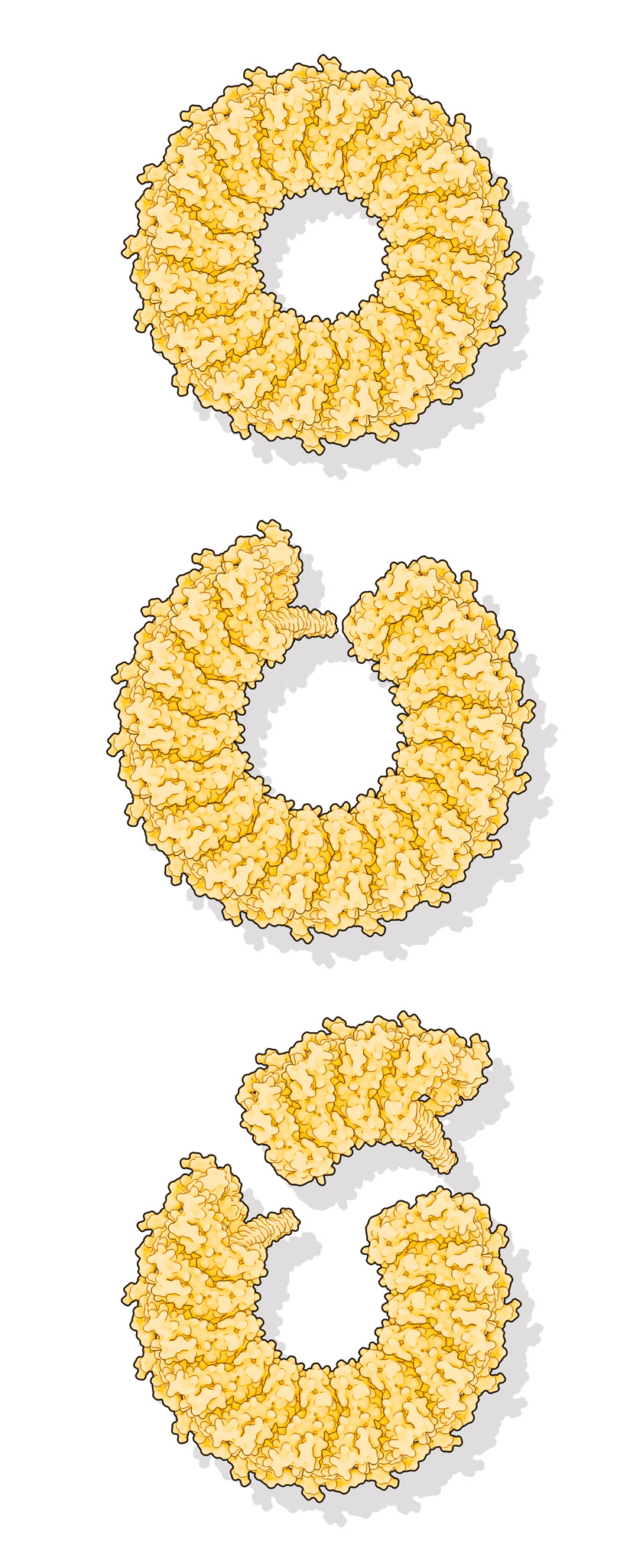
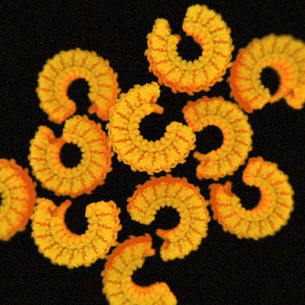
As its name suggests, the first one, perforin, pokes holes. Produced by the immune system to eliminate bacteria and infected or malignant cells, doughnut-shaped perforin molecules first land on their target. They then reconfigure to puncture that cell’s membrane, opening a passageway for toxic enzymes to finish the destruction of the target cell.

Cryo-electron microscopy studies had already determined the detailed structures for perforin just before and after it pokes through a membrane, but no one knew what happened in between. With HS-AFM, the group documented this transition, which traverses the round molecule section by section, like dominoes falling clockwise. It often stops before completing the circle. An incomplete ring appears adequate to do the job.
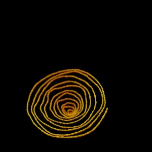
ESCRT
Microscopic image of yellow proteins in a spiral

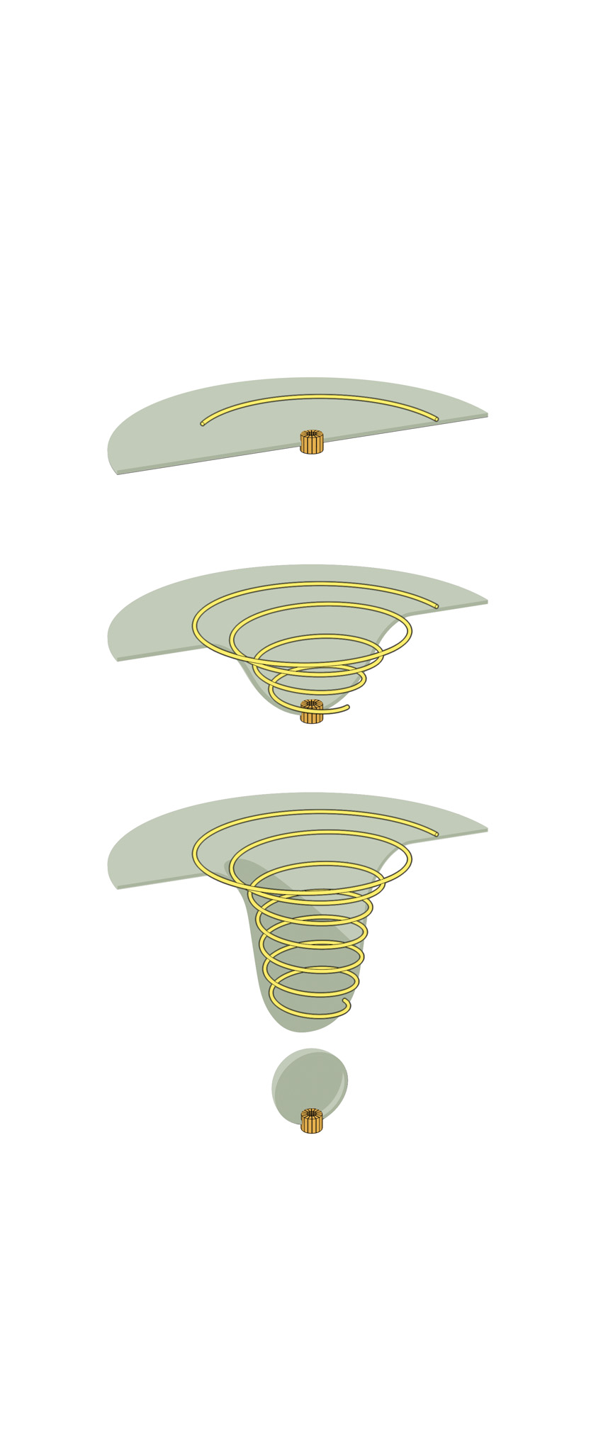
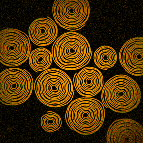
The second structure consists of elegant spirals built by the inelegantly named endosomal sorting complex required for transport system, otherwise known as ESCRT. As the name suggests, it helps cells sort and transport waste. ESCRT also enables them to sever connections and repair wounds.

HS-AFM recordings have tracked the formation of these structures, leading researchers to suggest they behave like springs, with the spiral compressing inward as the filament grows. Ultimately, the pressure forces its inner rings to pop outward.

Just as it protects the membranes of healthy cells, the spirals come to the aid of malignant ones. A study led by researchers with the company Genentech recently found that when under attack by perforin, tumor cells heal their wounds with ESCRT. This discovery suggests ESCRT could, perhaps, serve as a target for cancer drugs that aid the immune system. 


A Way In

There is a catch, however. Any therapy that disrupts these wound-healing spirals in cancer risks interfering with them in healthy cells. Should anyone follow up on the possibility of an ESCRT-jamming drug, HS-AFM would provide a straight-forward method to examine how it affects the formation of these spirals on the surface of malignant or normal cells, Dr. Scheuring says.

In general, membrane proteins have proven themselves immensely important to treating disease. They are estimated to account for the majority of molecules targeted by medications.

For Dr. Scheuring, this bias is intuitive. “In order to have an effect on our being, drugs must pass through the membrane barrier, generally by interacting with these proteins,” he says. “In some ways, all of what we are is inside cells.”

Illustration of cancer attacking a T-cell.

Summer 2023 Front to Back

  • From the Dean

    Message from the Dean

    As Weill Cornell Medicine marks the 125th year since its founding, it is striking to reflect upon how our values have endured.
  • Features

    Window Into the Future

    An ambitious research program could hold clues to improving the health of women and their children across their lifespans.
  • Features

    Caught on Camera

    Recordings made in Dr. Simon Scheuring’s lab reveal how elusive molecules embedded in cell membranes get their jobs done — for good and ill.
  • Features

    Risks and Rewards

    Alumnus Dr. Anthony Fauci (M.D. ’66) joins Dr. Jay Varma in a candid conversation about the future of public health and more.
  • Notable

    Two Landmark Anniversaries

    Weill Cornell Medicine is celebrating more than a century of excellence in medical education, scientific discovery and patient care, commemorating 125 years since its founding.
  • Notable

    Honoring Diversity

    In a celebration of Weill Cornell Medicine’s commitment to fostering diversity, equity and inclusion in academic medicine, the institution honored nearly a dozen faculty, students and staff.
  • Notable

    Overheard

    Weill Cornell Medicine faculty members are leading the conversation about important health issues across the country and around the world.
  • Notable

    News Briefs

    Notable faculty appointments, honors, awards and more — from around campus and beyond.
  • Notable

    Dateline

    The Salzburg-Cornell Seminars, now part of the Open Medical Institute (OMI), celebrates 30 years of knowledge-sharing, having served more than 26,000 fellows from 130 countries.
  • Grand Rounds

    There Is Hope

    How immunotherapy offered a new lease on life
  • Grand Rounds

    Medical School, Minus the Debt

    Weill Cornell Medicine’s debt-reduction program.
  • Grand Rounds

    News Briefs

    The latest on teaching, learning and patient-centered care.
  • Discovery

    Filling a Critical Gap in the Gut

    An important discovery positions fungi as a missing part of research on how the gut biome influences health.
  • Discovery

    New Clues to Coma Recovery

    Delays in regaining consciousness may serve a purpose: protecting the brain from oxygen deprivation.
  • Discovery

    Findings

    The latest advances in faculty research, published in the world’s leading journals.
  • Alumni

    Profiles

    Forging critical connections to move research from the bench to the bedside, our alumni are making an impact.
  • Alumni

    Notes

    What’s new with you? Keep your classmates up to date on all your latest achievements with an Alumni Note.
  • Alumni

    In Memoriam

    Marking the passing of our faculty and alumni.
  • Alumni

    Moments

    Marking celebratory events in the lives of our students, including Match Day and Graduation.
  • Second Opinion

    Nurturing Well-Being

    How can the health-care workforce recover from pandemic burnout?
  • Exchange

    High-Risk, High-Reward

    Two enterprising scientists discuss how the ecosystem for innovation at Weill Cornell Medicine provides the support entrepreneurial faculty and students need to turn their promising research into commercially viable drugs and other treatments.
  • Muse

    In the Flow

    Dr. Navarro Millán is a rheumatologist, clinical investigator and multi-instrumentalist.
  • Spotlight

    Making Health Care Affordable and Equitable

    Dr. Cheryl Pegus (M.D. ’88) is a cardiologist working in health-care businesses on new products to meet consumer needs, enhance health equity and improve health outcomes.